Human tooth
Article
Talk
Read
View source
View history
Tools
This is a good article. Click here for more information.
Page semi-protected
From Wikipedia, the free encyclopedia
For other uses of tooth or teeth, see Tooth (disambiguation).
Teeth
06-10-06smile.jpg
Image showing incisors and canine teeth, situated in gums above and below.
Human tooth diagram-en.svg
Diagram of a human molar showing its major constituents
Details
Identifiers
Latin dentes
TA98 A05.1.03.001
TA2 2818
FMA 75150
Anatomical terminology
[edit on Wikidata]
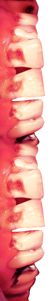
Human teeth function to mechanically break down items of food by cutting and crushing them in preparation for swallowing and digesting. As such, they are considered part of the human digestive system.[1] Humans have four types of teeth: incisors, canines, premolars, and molars, which each have a specific function. The incisors cut the food, the canines tear the food and the molars and premolars crush the food. The roots of teeth are embedded in the maxilla (upper jaw) or the mandible (lower jaw) and are covered by gums. Teeth are made of multiple tissues of varying density and hardness.
Humans, like most other mammals, are diphyodont, meaning that they develop two sets of teeth. The first set, deciduous teeth, also called "primary teeth", "baby teeth", or "milk teeth", normally eventually contains 20 teeth. Primary teeth typically start to appear ("erupt") around six months of age and this may be distracting and/or painful for the infant. However, some babies are born with one or more visible teeth, known as neonatal teeth or "natal teeth".
Anatomy
Main article: Dental anatomy
There are four main types of teeth in humans, shown labelled here.
Dental anatomy is a field of anatomy dedicated to the study of tooth structure. The development, appearance, and classification of teeth fall within its field of study, though dental occlusion, or contact between teeth, does not. Dental anatomy is also a taxonomic science as it is concerned with the naming of teeth and their structures. This information serves a practical purpose for dentists, enabling them to easily identify and describe teeth and structures during treatment.
The anatomic crown of a tooth is the area covered in enamel above the cementoenamel junction (CEJ) or "neck" of the tooth.[2][3] Most of the crown is composed of dentin ("dentine" in British English) with the pulp chamber inside.[4] The crown is within bone before eruption.[5] After eruption, it is almost always visible. The anatomic root is found below the CEJ and is covered with cementum. As with the crown, dentin composes most of the root, which normally has pulp canals. Canines and most premolars, except for maxillary first premolars, usually have one root. Maxillary first premolars and mandibular molars usually have two roots. Maxillary molars usually have three roots. Additional roots are referred to as supernumerary roots.
Humans usually have 20 primary (deciduous, "baby" or "milk") teeth and 32 permanent (adult) teeth. Teeth are classified as incisors, canines, premolars (also called bicuspids), and molars. Incisors are primarily used for cutting, canines are for tearing, and molars serve for grinding.
Most teeth have identifiable features that distinguish them from others. There are several different notation systems to refer to a specific tooth. The three most common systems are the FDI World Dental Federation notation (ISO 3950), the Universal Numbering System, and the Palmer notation. The FDI system is used worldwide, the Universal only in the United States, while the older Palmer notation still has some adherents only in the United Kingdom.
Primary teeth
Among deciduous (primary) teeth, ten are found in the maxilla (upper jaw) and ten in the mandible (lower jaw), for a total of 20. The dental formula for primary teeth in humans is
2.1.0.2
2.1.0.2
.
In the primary set of teeth, in addition to the canines, there are two types of incisors—centrals and laterals—and two types of molars—first and second. All primary teeth are normally later replaced with their permanent counterparts.
The Universal Numbering System for adult human teeth. The view is from a dental practitioner's perspective, meaning tooth 1 is the upper right rear (third) molar.
Permanent teeth
Among permanent teeth, 16 are found in the maxilla and 16 in the mandible, for a total of 32. The dental formula is
2.1.2.3
2.1.2.3
. Permanent human teeth are numbered in a boustrophedonic sequence.
The maxillary teeth are the maxillary central incisors (teeth 8 and 9 in the diagram), maxillary lateral incisors (7 and 10), maxillary canines (6 and 11), maxillary first premolars (5 and 12), maxillary second premolars (4 and 13), maxillary first molars (3 and 14), maxillary second molars (2 and 15), and maxillary third molars (1 and 16). The mandibular teeth are the mandibular central incisors (24 and 25), mandibular lateral incisors (23 and 26), mandibular canines (22 and 27), mandibular first premolars (21 and 28), mandibular second premolars (20 and 29), mandibular first molars (19 and 30), mandibular second molars (18 and 31), and mandibular third molars (17 and 32). Third molars are commonly called "wisdom teeth" usually emerge at ages 17 to 25.[6] These molars may never erupt into the mouth or form at all.[citation needed] When they do form, they often must be removed. If any additional teeth form—for example, fourth and fifth molars, which are rare—they are referred to as supernumerary teeth (hyperdontia). Development of fewer than the usual number of teeth is called hypodontia.
There are small differences between the teeth of males and females, with male teeth along with the male jaw tending to be larger on average than female teeth and jaw. There are also differences in the internal dental tissue proportions, with male teeth consisting of proportionately more dentine while female teeth have proportionately more enamel.[7]
Parts
Human tooth diagram-en.svg
Enamel
Main article: Tooth enamel
Enamel is the hardest and most highly mineralized substance of the body. It has its origin from oral ectoderm. It is one of the four major tissues which make up the tooth, along with dentin, cementum, and dental pulp.[8] It is normally visible and must be supported by underlying dentin. 96% of enamel consists of mineral, with water and organic material comprising the rest.[9] The normal color of enamel varies from light yellow to grayish white. At the edges of teeth where there is no dentin underlying the enamel, the color sometimes has a slightly blue tone. Since enamel is semitranslucent, the color of dentin and any restorative dental material underneath the enamel strongly affects the appearance of a tooth. Enamel varies in thickness over the surface of the tooth and is often thickest at the cusp, up to 2.5mm, and thinnest at its border, which is seen clinically as the CEJ.[10] The wear rate of enamel, called attrition, is 8 micrometers a year from normal factors.[11]
Enamel's primary mineral is hydroxyapatite, which is a crystalline calcium phosphate.[12] The large amount of minerals in enamel accounts not only for its strength but also for its brittleness.[10] Dentin, which is less mineralized and less brittle, compensates for enamel and is necessary as a support.[12] Unlike dentin and bone, enamel does not contain collagen. Proteins of note in the development of enamel are ameloblastins, amelogenins, enamelins and tuftelins. It is believed that they aid in the development of enamel by serving as framework support, among other functions.[13] In rare circumstances enamel can fail to form, leaving the underlying dentine exposed on the surface.[14]